
Health Tech
Ultrasound patches go cable-free
Wearable ultrasound patches can revolutionise health care, but until now have relied on cables linked to a device.
Wearable ultrasound patches have the potential to revolutionise health care, facilitating the remote monitoring of critical physiological functions in the comfort of a patient’s home. But most patches in development have a major limitation: they require cables to power the device and transmit the ultrasound data, physically tethering the wearer to a control system. That is, until now.
A fully wireless ultrasound patch that can continuously track critical vital signals such as heart rate and blood pressure was recently reported in Nature Biotechnology. The patch, which can capture detailed medical information and wirelessly transmit the data to a smart device (such as a laptop or smartphone), could represent a major step forward in at-home health care technology.
“The true impact of wearable ultrasound patches has yet to be fully realised, as previously described iterations aren’t wireless and limit the users’ ability to go about their daily lives,” noted Randy King, Ph.D., a program director in the Division of Applied Science & Technology at NIBIB. “The technology described here represents necessary and essential progress in the wearable ultrasound space, potentially unlocking the promise of remote ultrasound monitoring for any number of health conditions.”
Ultrasound, which uses sound waves and their resulting echoes to image tissues inside the body, has traditionally been limited to the clinic. The ultrasound patch technology in this study was pioneered by Sheng Xu, Ph.D., an associate professor and Jacobs Faculty Scholar at the University of California San Diego (UC San Diego). His team has previously reported wearable ultrasound patches with similar transducers. The real advancement in this latest study is the wireless capability of the patch.

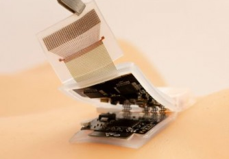
“The key element of this study is the design of the ultrasound circuit,” said Xu. “In our previous patches, the ultrasound probe was connected to a flexible cable for power and data transmission. In this patch, the cables are replaced with a wearable circuit, which can pre-process and wirelessly transmit the ultrasound data to a back-end station for further analysis.”
The ultrasound system is composed of a probe, a circuit, and a battery. As the current system was designed with a focus on cardiovascular health, the ultrasound probe was typically placed on the carotid artery in this study. The probe is attached to a flexible circuit, which activates the ultrasound transducers, collects the ultrasound echoes, amplifies and filters these echoes, and transmits the digitised signal to a terminal device. The entire system is powered by a commercial rechargeable lithium polymer battery.
“We developed a machine learning algorithm to coordinate with the circuit to automatically process the ultrasound signals and continuously track the carotid artery, which allows us to obtain ultrasound information even when the wearer of the patch is moving,” explained first author Muyang Lin, a Ph.D. candidate in the Xu laboratory. “This automatic tracking algorithm provides unprecedented opportunities for medical ultrasonography and exercise physiology.”
To assess its generalizability, the researchers cross-validated their machine learning model among ten healthy subjects representing three distinct racial groups. Using a machine learning technique called domain adaptation, the researchers found that a model trained on data from one participant wearing the patch could be successfully adapted for the other participants. With minimal model retraining, the patch could track the pulsations of the carotid artery with high accuracy, allowing for measurements like blood pressure, arterial stiffness, and cardiac output. Continual monitoring of these measurements among high-risk populations could provide advance warning of heart failure.
“Validating our patch in a larger population is a crucial next step,” said Lin. “We are working on validating our sensor against existing medical devices.”
While the device was mainly evaluated on its ability to monitor cardiovascular functions, the researchers also demonstrated that the patch can be applied to the abdomen for diaphragm monitoring or to limbs for peripheral artery monitoring. “The system holds the potential to perform measurements at multiple spots in the body, and we can easily tailor the probe design to fit diverse tissue monitoring requirements,” said Xu.
“With this kind of device, we hope to blur the boundary between at-home care and in-hospital diagnosis,” said Lin. “We foresee a future where diagnoses can occur anytime and anywhere, enabled by wireless devices like these.”


