



Health Tech
Philips maps AI’s route through healthcare
A visit to the Philips Customer Experience Centre in the Netherlands revealed a broad range of AI-powered medical technologies, writes JASON BANNIER.

Philips is pushing healthcare AI into practical tools that change how scans, screenings and patient monitoring are performed.
During a visit to the Philips Customer Experience Centre in the Netherlands last week, the showcased technologies pointed to ways AI could ease healthcare workloads, shorten examinations and bring screening closer to patients.
The demonstrations included a handheld pregnancy ultrasound system, AI-assisted cardiac MRI, AI-supported CT imaging and a wearable heart monitor that can be posted to a patient’s home.
A central theme ran through all of them: AI is being used less as a replacement for clinicians and more as a way to guide people through complex medical processes, automate repetitive tasks, and identify patients who need specialist attention.
One of the clearest examples was SmartSweep, an AI-powered ultrasound system intended for pregnancy screening in areas where access to trained sonographers is limited.
The system combines a wired handheld ultrasound probe with an Android tablet. The application guides the user through the examination step by step, asking for basic patient information, then instructing the operator to perform vertical and horizontal sweeps across the abdomen. The probe can fit inside a backpack or handbag, while all processing runs locally on the tablet, without requiring an internet connection.
“Our goal isn’t simply to generate an ultrasound image,” said Jeroen Maas, Philips director for access to care technology and partnerships. “Our goal is to provide the right clinical information while solving the shortage of trained medical personnel.”
During the demonstration, Maas scanned a pregnancy training model containing a simulated 20- to 21-week foetus. The software coached each sweep in real time, rejecting movements when the probe was tilted or moved incorrectly, and accepting the sweep only once the operator corrected the angle.

The aim is not to replace a full diagnostic ultrasound. SmartSweep functions as a screening tool that can estimate gestational age, calculate the expected delivery date, identify a possible multiple pregnancy, assess foetal cardiac activity, evaluate foetal presentation where relevant, and flag possible problems, such as abnormal amniotic fluid levels. Patients with alerts can then be referred for a comprehensive ultrasound examination by a qualified professional.
The system is being clinically evaluated in Kenya and India, with several African countries also showing interest. Philips says the product is being prepared for commercial release, subject to local regulatory approvals.
In a separate demonstration, Gwen Herigault, Philips head of clinical programmes for MR, showed how AI is changing cardiac MRI, a field that has traditionally required long scan times and highly specialised staff.
Cardiac MRI can provide detailed images of the heart muscle, but limited scanner capacity, examination complexity and staff shortages have restricted wider use. Philips says AI and automation can now reduce a cardiac MRI examination that previously took about an hour to around 15 minutes.

The company’s SmartHeart workflow automates much of the planning required before a cardiac scan. According to the demonstration, a process that could previously require around 130 mouse clicks can be reduced to three, with the technologist still checking that the automated planning is correct.
“The goal isn’t simply automation,” said Herigault. “The goal is autonomy.”
AI can support faster image acquisition, with some scan sequences falling from roughly one minute to about ten seconds. Patient setup has been simplified as well, with camera-based heart-rate detection replacing the need for electrocardiogram (ECG) electrodes in some workflows.
Herigault said autonomous systems do not remove the healthcare professional from the process.
“The technology performs more of the work, but the healthcare professional always remains responsible.”
Philips highlighted helium-free MRI systems as part of the access story. Traditional MRI scanners require liquid helium and more complex installation conditions. The newer systems are designed to give hospitals greater flexibility in where scanners can be installed, including mobile trucks and community healthcare environments.
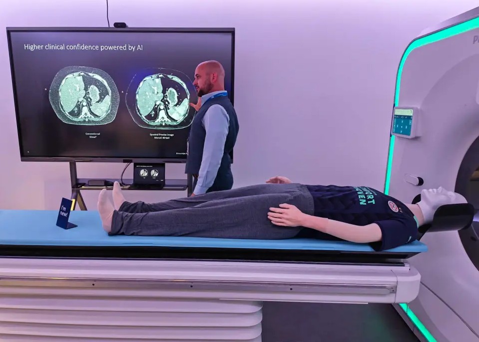
A CT scanner demonstration focused on a different pressure point: patient volume.
Bas Stam, Philips business development manager for CT and AMI, described CT as the “backbone” of a radiology department because the modality supports neurological, stroke, cardiac, oncology, trauma and other examinations.
CT scans are fast. A chest, abdomen and pelvis scan, or even a full-body scan, can take between two and six seconds. However, some hospitals scan far more patients per machine than others, with high-volume regions reaching 150 to 250 patients per scanner per day. That creates a need for more autonomous workflows.
“So, when we look at the transformation of CT services through AI, we’re asking one central question,” said Stam. “How can we create enough capacity to scan even more patients?”
Philips uses AI across the CT workflow, from patient positioning to image reconstruction and post-processing. A ceiling-mounted camera, referred to as Precise Positioning, helps place the patient more consistently, reducing reliance on manual positioning by radiographers.

Deep-learning image reconstruction can improve CT image quality from the same raw scan data, rather than increasing radiation dose. The company says that helps sharpen images, reduce noise and improve contrast while maintaining focus on dose reduction.
Automation can play a role in supporting post-processing. In trauma cases, tools such as Precise Rib can automatically identify and label ribs within seconds after image reconstruction begins, while other workflows can standardise brain scan orientation when a patient cannot be positioned perfectly.
Philips presented Spectral CT as a step beyond conventional anatomical imaging. The technology can help identify materials such as iodine and calcium, giving clinicians more quantitative information about what they are seeing, not only where a structure is located.
The most consumer-like device shown during the visit was ePatch, a small wearable heart monitor used to detect arrhythmias such as atrial fibrillation. The patch can be worn for up to 14 days and works with Philips’ Cardiologs AI platform to help clinicians identify clinically significant heart rhythm events from large volumes of ECG data. Gadget featured the device this week as a Product of the Day for helping doctors spot irregular heart rhythms that short hospital tests may miss.
The visit showed us that healthcare AI is becoming most useful when the technology disappears into workflow: a progress bar guiding a pregnancy scan, a camera helping position a CT patient, software planning an MRI exam, or an algorithm sorting through days of heart rhythm data.
Philips’ live procedure technology, seen separately by Gadget at St Antonius Hospital, applies the same principle in the operating room, where AI-guided imaging helps doctors navigate cardiac repair devices inside the beating heart during minimally invasive procedures. The full story will appear as Gadget’s lead article on Monday (13 July 2026).
- See Gadget’s report on Philips’ Future Health Index 2026, which found that clinicians are adopting AI faster than many health systems can support the technology safely and consistently, here.
* Jason Bannier is a data analyst at World Wide Worx and deputy editor of Gadget.co.za. Follow him on Bluesky at @jas2bann.